General Information about the Knee
Anatomy of the Knee
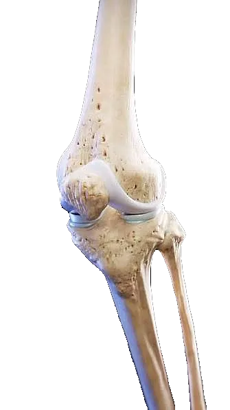
The knee is a joint that connects the thigh to the lower leg and is formed by the distal end of the femur, the proximal end of the tibia, the patella, and the surrounding soft tissues. The patellofemoral joint is formed between the femur and the patella, and the femorotibial joint is formed between the femur and the tibia.
In healthy knees, the ends of the bones are covered by cartilage, a cushioned, elastic tissue that prevents direct friction between the bones. This allows for free, painless movement at the joint. Muscles and ligaments provide mobility and stability, respectively, to the knee, and synovial tissues produce fluid that acts as a lubricant for the joint.
The knee is one of the joints in the human skeleton most commonly affected by osteoarthritis. The reason for the high prevalence of knee osteoarthritis, compared to other joints in the body, is that the knee is a “weight-bearing” joint; that is, it must support the body’s weight and the weight of the objects we carry when we are standing, or when we move by walking, running, or going up or down stairs.
Knee Conditions
The main knee disorders and conditions can be grouped according to their cause:
1. Advanced Degenerative Disease (Joint Replacement Surgery)
- Severe knee osteoarthritis (gonarthrosis): When the cartilage has worn away completely and there is direct bone-on-bone contact. The surgeon performs a knee replacement (implantation of a total or partial knee prosthesis).
Axial Deformities (Genu Varo / Genu Valgo): “Bowlegs” or “knock-knees” that cause uneven wear. In young patients with osteoarthritis on only one side, the surgeon may perform an osteotomy (cutting and realigning the bone) to delay the need for a prosthesis by several years.
Previous Prosthesis Failures and Loosening: Complex Prosthesis Revision Surgeries When Old Components Have Worn Out, Become Loose, or Become Infected.
2. Sports Injuries
Cruciate ligament tears (ACL and PCL): The surgeon performs a ligament reconstruction using a graft (from the patient or from a tissue bank).
Complex or unstable meniscal tears: Especially those that can be sutured (repaired) rather than removed, to protect the cartilage in the long term. If the meniscus is severely damaged, meniscal transplants may be performed.
Osteochondral (cartilage) injuries: Holes or “defects” in the cartilage caused by trauma. The surgeon uses regeneration or repair techniques, such as microfractures, mosaicplasty (cartilage graft), or chondrocyte implantation.
3. Patellar Instability and Disorders
Recurrent patellar dislocation: Patients whose patella repeatedly “pops out of place.” The surgeon performs surgery to reconstruct the medial femoropatellar ligament (MFP) or realign the bone (tibial tuberosity) so that the patella glides properly.
Severe patellar hyperpressure syndrome: Severe cases in which the patella exerts excessive, asymmetric pressure on the femur, requiring surgical release of the lateral retinaculum or realignment.
4. Trauma and Emergency Care
Complex fractures: Fractures of the tibial plateau, the patella, or the femoral condyles.
Joint infections (septic arthritis): A medical emergency that requires immediate surgical irrigation of the joint to prevent bacteria from destroying the cartilage within a matter of hours.
Learn More